

In this article, we will discuss What is Neuron? Describe the Structure of a Neuron. Explain the Process of Neural Conduction.
What is Neuron?
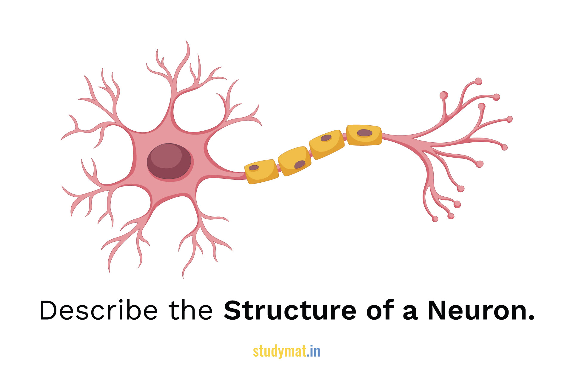
The cells of the Nervous System that receive and send messages within that system are called Neurons. Neurons receive and process information to and from the brain. Nerve Impulse is the electrical signal that the neuron conduct. They are in differing shapes and sizes having diverse functions to perform. Though, the neuron structure is related to its functions. Here is an illustration of the external structures of a type of Neuron.

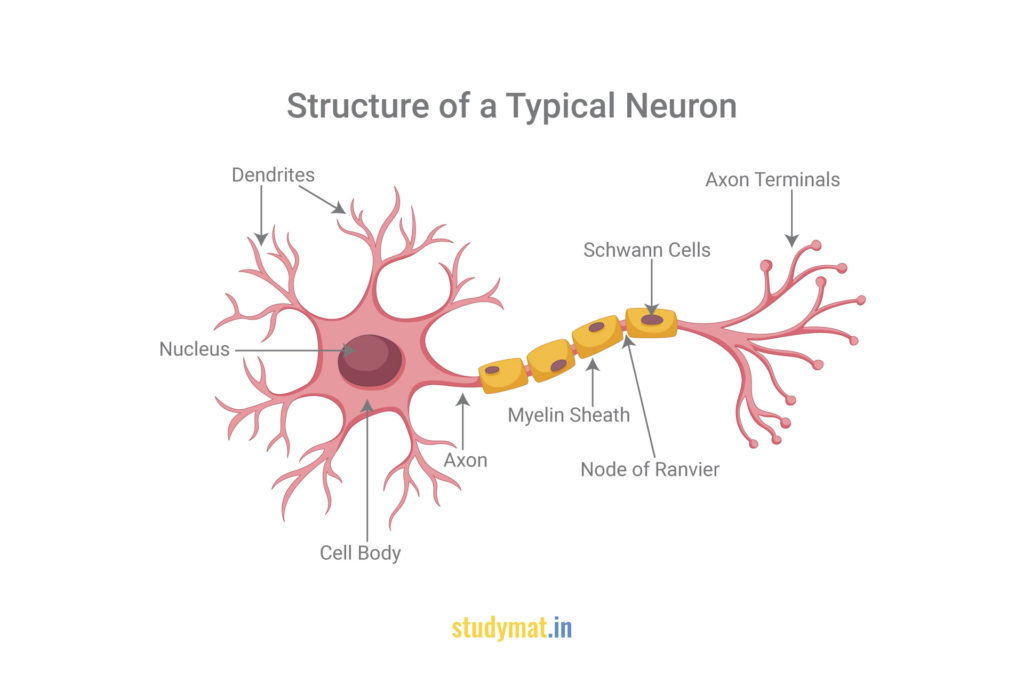
Structure of a Neuron.
Neuron has the cell body, axon, and terminal buttons. The plasma membrane of the neuron is the double layer of phospholipid molecules. It is semipermeable to certain kinds of substances. The Plasma Membrane controls the movement of the substances through it. Consequently, it involves the nerve impulse. It also provides sites for the electrical activity that happens during nerve impulses and for synaptic activity between two neurons.
Cell Body, Nucleus, Dendrites, Axon, Axon Hillock, Terminal Buttons, Myelin Sheath, and Axon Collaterals:
- The Cell Body is the largest part of the Neuron. It has some organelles floating in its cytoplasm like the Golgi body, Mitochondria, Nissl bodies, Smooth Endoplasmic Reticulum, Rough Endoplasmic Reticulum, etc. Most importantly, the Cell Body is the metabolic center of the Neuron and is also known as Soma (means body).
- There is a Nucleus in the centre. The Dendrites are the extensions from the cell body, it look like branches of a tree. The dendrite tips have sensory receptors which receive the stimuli from other neurons. It begins the process of the nerve impulse by sending these impulses to the Cell Body.
- The Axon is a long slender part of the neuron which extends from a portion of the cell body called the Axon Hillock.
- Axon Hillock is a cone-shaped region at the connection between the axon and the cell body.
- It is frequently covered by the Myelin Sheath and conveys information from the cell body towards its distal ends called Terminal Buttons.
- Myelin Sheath is fatty insulation around many axons. The axons differ in length and diameter. Larger diameters indicating faster action potentials, influencing neural conduction.
- The axon may have branches called Axon Collaterals.
An electrical message or brief chemical that begins from the cell body travels down the axon to the terminal buttons is called an Action Potential. When the axons divide at the rear end, it branches profusely and each branch ends in a Knob. These are called Terminal Buttons. When the action potential travels down the axon, it reaches the terminal buttons, where a chemical substance is released called the Neurotransmitter. The function of the neurotransmitters is to either increase or decrease the activity of the receiving neuron.
However, Neurons are present in large numbers in the brain, there are other primary cells that provide support to the neurons, called Neuroglia, Glial Cells, or Glia. Glial Cells affect thinking, memory, learning, perception, and help in maintaining a state of Homeostasis of the Nervous System.
Glial Cells:
This Cells deliver nutrients to neurons, produce myelin to coat axons, help in information processing, clean up waste products and dead neurons, and influence the generation of new neurons throughout prenatal development. Meanwhile, Neuroscientists investigated the role of Glial Cells in neurodevelopmental disorders, such as Autism Spectrum Disorder, degenerative disorders, such as Alzheimer’s, and psychiatric disorders such as Schizophrenia and Major Depressive Disorder.
There are four types of Glial Cells. They are Oligodendrocytes, Schwann Cells, Microglia, and Astrocytes. Oligodendrocytes produce myelin for neurons in the brain and spinal cord or the central nervous system. Schwann Cells produce myelin sheath around the neurons of the body or the peripheral nervous system. Myelin protects the shaft of the axon as well as gives support. These nerve fibers are called Myelinated Fibers. There are certain gaps between the myelin sheath which are called Nodes of Ranvier. Microglial Cells are involved in inflammation response, which is protecting the brain from invading microorganisms. Astrocytes clean up the waste material of dying neurons.
Follow us:
If you like this article, you can Follow us on Facebook.
Also, you can Join our Official Facebook Group for QnA Sessions and Discussions with the worldwide IGNOU community.